29
Few phrases in modern healthcare carry the unearned cultural dread of a root canal. In popular media and casual conversation, the procedure is routinely invoked as the ultimate benchmark for discomfort. Yet this widespread reputation is decades out of date. Modern endodontic therapy does not cause pain; it relieves it. In reality, a root canal is a sophisticated, tooth-preserving procedure that clears deep bacterial infection, stops progressive bone loss, and saves a natural tooth that would otherwise require surgical extraction.
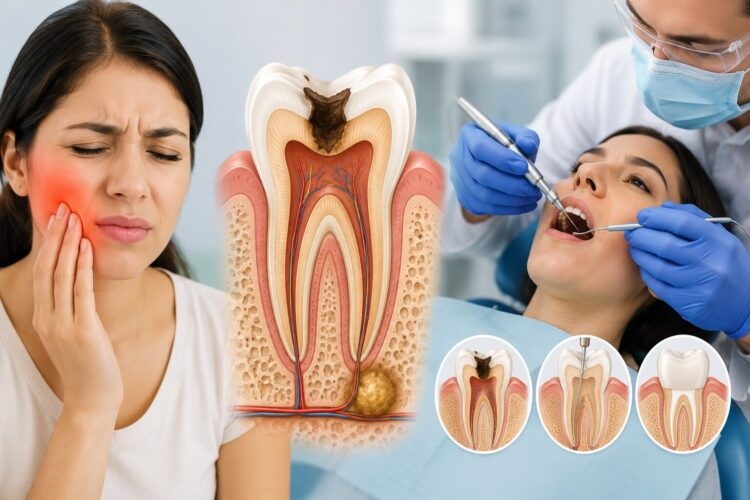
The human tooth is not a solid stone. Beneath the protective outer shields of hard enamel and yellow dentin lies a delicate, hollow chamber housing the dental pulp. This pulp contains a living network of blood vessels, specialized connective tissue, and sensitive nerve fibers responsible for nourishing the tooth during its developmental stages. When a deep cavity, an invisible hairline crack, or blunt trauma breaches that protective barrier, oral bacteria invade the pulp chamber. Once infected, the pulp cannot heal on its own. Recognizing the subtle and overt distress signals of an infected pulp is the critical first step in intervening before a localized infection transforms into a severe facial abscess.
Recognizing the Warning Signs of Deep Pulp Infection
The human dental nerve communicates primarily through pain, but the character, duration, and triggers of that discomfort provide vital diagnostic clues about what is happening beneath the gumline.
Persistent, Spontaneous Throbbing
A brief twinge of discomfort while chewing a tough piece of food is not necessarily an endodontic emergency. However, pain that is spontaneous, unprovoked, and persistent points toward irreversible inflammation of the dental pulp. If a tooth begins to throb relentlessly while you are resting on the couch, or wakes you from a sound sleep in the middle of the night, the internal nerve is likely suffocating from mounting pressure within the rigid pulp chamber. This pain often radiates outward, confusing the brain into perceiving discomfort in the jawbone, cheek, ear, or neighboring teeth.
Lingering Thermal Sensitivity
Almost everyone has experienced momentary sensitivity when sipping an icy drink or biting into hot soup. In a healthy tooth, that sensation vanishes the second the hot or cold stimulus is removed. Lingering thermal sensitivity is a primary indicator of irreversible pulpitis. If drinking hot coffee triggers a sharp or dull ache that hangs around for thirty seconds, two minutes, or even longer after swallowing, the nerve tissue has suffered severe cellular damage. Sensitivity to heat is particularly telling, as it often suggests the pulp has begun to decompose, producing expanding gases inside the tooth.
Tenderness to Touch and Chewing Pressure
When an infection consumes the entire pulp chamber, bacteria migrate through the microscopic opening at the tip of the tooth root, known as the apical foramen. Once bacteria escape into the surrounding jawbone, they inflame the periodontal ligament—the sling of fibrous tissue that anchors the tooth in the bone socket. When this occurs, biting down, tapping the tooth with a fingernail, or even touching it with your tongue generates a bruised, sharp tenderness. If you instinctively chew on one side of your mouth to avoid touching a specific tooth, the structural support surrounding that root is likely inflamed.
Gum Pimples, Swelling, and Drainage
The emergence of a small, pimple-like bump on the gums adjacent to a painful tooth—clinically known as a fistula or sinus tract—is an unmistakable sign of an active infection. As white blood cells battle bacteria at the root tip, pus accumulates. The body attempts to relieve this internal pressure by creating a microscopic drainage tunnel through the jawbone and soft gum tissue. You may notice an unpleasant, metallic taste in your mouth or an intermittent foul odor as the abscess drains. While this drainage temporarily relieves pressure and reduces pain, the underlying infection remains fully active.
Deep Tooth Discoloration
A tooth that gradually darkens, shifting to a dull gray, slate, or brownish hue, is often signaling internal trauma. Much like a bruise under a fingernail, deep tooth discoloration frequently occurs when injured pulp tissue suffers internal bleeding or complete necrosis. Even if you cannot recall hitting the tooth, sports trauma or an impact from years ago can quietly disrupt the blood supply, causing the nerve to die slowly over a long period.
The Silent Hazard: When the Pain Suddenly Stops
One of the most dangerous developments in dental health is the abrupt disappearance of severe toothache pain without clinical treatment. Patients often assume that because the throbbing has stopped, the underlying issue has resolved itself.
In reality, the cessation of pain usually means that the sensory nerve fibers inside the tooth have completely died. The bacteria, however, have not vanished. With the nerve tissue dead, bacterial colonies multiply unchecked inside the hollow, dark root canals, steadily eroding the surrounding jawbone. Left untreated, a dormant infection can erupt into a systemic cellulitis, causing dangerous facial swelling that requires emergency hospitalization. An asymptomatic infected tooth is a ticking clock; the absence of pain does not equal the absence of disease.
Demystifying the Procedure: What to Expect in the Chair
Understanding the methodical steps of modern endodontic therapy dispels the mystery and anxiety surrounding the appointment. The entire procedure is designed around comfort, microscopic precision, and clinical sterility.
Profound Local Anesthesia and Isolation
Before any treatment begins, the clinician administers targeted local anesthesia to numb the tooth and surrounding tissues completely. With modern numbing agents, the sensation during a root canal is no different than having an ordinary filling placed. Once profound numbness is confirmed, the dentist places a thin sheet of rubber or silicone called a dental dam over the tooth. This dam isolates the tooth from the rest of the mouth, keeping the operational field completely dry, sterile, and protected from salivary bacteria.
Canal Disinfection and Shaping
The practitioner creates a small access opening through the biting surface of a molar or the backside of a front tooth to expose the pulp chamber. Using specialized magnification, such as high-powered surgical loupes or an operating microscope, the doctor locates the tiny canal entrances.
Using ultra-flexible, rotary nickel-titanium instruments, the clinician gently removes the diseased pulp tissue, cleans the intricate walls of the canals, and shapes them into smooth, tapered corridors. Throughout this mechanical shaping, the doctor floods the canals with antimicrobial irrigating solutions to dissolve lingering bacteria, necrotic debris, and organic tissue from hidden microscopic crevices.
Hermetic Sealing
Once the canals are meticulously cleaned, disinfected, and dried with sterile paper points, they must be permanently sealed to prevent future bacterial recolonization. The doctor fills the clean internal root canals with a biocompatible, rubber-like material derived from tree resin called gutta-percha. Paired with an adhesive sealer paste, the gutta-percha is condensed into place to create a hermetic seal from the crown down to the apex of the root. The access cavity is then sealed with a temporary or permanent restorative filling.
The Essential Final Phase: Permanent Structural Restoration
Completing the root canal cleans out the infection, but it does not finish the job of preserving the tooth. A tooth that has undergone endodontic therapy loses its internal vascular hydration, making the remaining enamel and dentin significantly more brittle over time. Furthermore, the combination of prior decay, old fillings, and the access opening leaves the physical walls of the tooth structurally compromised.
For posterior teeth—the premolars and molars that absorb hundreds of pounds of daily chewing force—a full-coverage dental crown is virtually mandatory. Placing a porcelain, zirconia, or metal-ceramic crown protects the remaining tooth structure like an architectural helmet, redistributing bite forces and preventing catastrophic vertical root fractures that would require an extraction. In most cases, patients return to their general dentist within a few weeks of root canal completion to have this final crown cemented into place.
Post-Treatment Recovery and Long-Term Prognosis
Recovery from a root canal is typically uneventful. For two to four days following treatment, the surrounding gum tissue and jaw muscles may feel mildly tender or bruised, particularly when chewing on that side. This temporary inflammation is easily managed with common over-the-counter anti-inflammatory medications like ibuprofen or acetaminophen. Most patients return to work, school, and normal daily activities the very next day.
When executed with modern techniques and protected by a durable crown, a root-canal-treated tooth boasts a clinical success rate well above ninety percent. It can remain fully functional, pain-free, and stable for decades—often for the rest of your life.
Preserving your natural teeth is always the gold standard of oral health. No bridge, removable partial denture, or dental implant perfectly replicates the natural tactile feedback and biomechanical efficiency of your original tooth structure. By paying attention to early warning signs and seeking prompt professional care, you turn what could be a painful dental emergency into a routine, comfortable procedure that keeps your natural smile intact.
